
Our goal is to make ECG interpretation simple, practical, and fun for acute providers; and we believe that mastery of this simple skill will save lives.
Joined September 2017
- Tweets569
- Following4
- Followers21.7K
- Likes24








Electrical alternans: the QRS changes size beat to beat. 🫀
In T-wave alternans, what changes from one beat to the next?
81%The T-wave size
19%The QRS axis
0%The PR interval
0%The heart rate
16 votes • Final resultsMYTH VS. REALITY? 🤔
Sgarbossa and modified Sgarbossa are accepted in the 2022 ACC/AHA chest pain guidelines.
24%Myth
76%Reality
33 votes • 22 hours
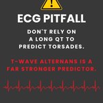
T-wave alternans can be an ominous warning: bigger, smaller, bigger, smaller from beat to beat. ⚠️
Dr. Mattu explains why this pattern deserves serious attention when you’re worried about impending torsades.
Would you recognize it in time?
ecgweekly.com/weekly-workout…
TRUE OR FALSE? 🤔
The Aslanger pattern is part of the formal ACC/AHA STEMI guidelines.
28%TRUE
72%FALSE
36 votes • Final resultsA 45 yo m presents to the emergency department with palpitations. He has no known history of coronary artery disease. He is alert, with a systolic blood pressure of approximately 120 mm Hg and a heart rate near 180 beats/min. The following ECG shows.......ecgweekly.com/weekly-workout…

ST elevation in lead III. None in II or aVF. 🫀
The computer reads it as nonspecific.
Why might it miss this?
73%Needs 2 contiguous leads
3%Lead III is unreliable
3%The rate is too fast
22%ST change is too small
37 votes • Final results
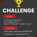
Chest pain. ST elevation in lead III only. ST in V1 higher than V2. Lateral ST depression with upright T waves. 🔎
Computer: nonspecific.
Nonspecific—or acute occlusion?

71M with chest pain. Lead III has ST elevation—but II and aVF don’t. 👀
The computer called it nonspecific. Cardiology: not a STEMI.
But the ED physician recognized a subtle high-risk pattern.
What did he see?
ecgweekly.com/weekly-workout…
MYTH OR REALITY? 👀
Any irregular rhythm without obvious P waves is atrial fibrillation.
61%MYTH
39%REALITY
41 votes • Final resultsA 76-year-old man presents with chest pain suggestive of acute coronary syndrome. An initial 12-lead ECG is obtained and shown below, but does not meet conventional STEMI criteria......ecgweekly.com/weekly-workout…

Grouped beats ≠ automatically AFib. 🔎
Map the P waves:
Regular → Mobitz
Premature → PACs
The rhythm may be hiding in the atria.

The computer says AFib—but the beats occur in repeating groups. 🔎
What should you map next?
The P waves.
Regular → think Mobitz
Premature → think PACs

📚 Last day of #PrimeDay!
Looking to build your ECG library? We've put together our recommended ECG books for every stage of learning—from beginners to advanced clinicians.
Learn why we recommend each one:
ecgweekly.com/ecgstat/book-r…
#AmazonAssociate #ECGWeekly #EmergencyMedicine
