

Become a better endoscopist with EndoCollab, the world's most comprehensive endoscopy community. Join us: https://xitter.cf/t.co/EkRtunpQyB
🌎
Joined February 2021
- Tweets10.2K
- Following2.9K
- Followers31.2K
- Likes21.2K




EndoCollab Quiz
"Funny Gastric Polyp"
- Polypoid lesion with depressed center
- Location: Antrum, Greater Curvature, Pre-Pyloric Area
Question: Most likely diagnosis?
Reply below with your answer.

EndoCollab™ | GI Endoscopy Community retweeted

Round, yellowish, submucosal. A: carcinoid. B: granular cell tumor, much less common.
In the rectum, a lesion like A is carcinoid until proven otherwise, not a lipoma. Injection: poor lift. EMR or ESD, never plain snare.
By Klaus Mönkemüller, MD, PhD
Migrated EndoBarrier: bleeding, outlet obstruction, pancreatitis. Barbs anchored in the distal duodenum.
80 cm overtube, pediatric colonoscope, hook on the metal crown, all metal inside the overtube before pulling.
Your approach?
Full lecture on EndoCollab: endocollab.com/blogs/full-co…

EndoCollab™ | GI Endoscopy Community retweeted
Granular cell tumors can be malignant from the outset. We favor resecting by histology (Fanburg-Smith), not symptoms alone. Case by Klaus Mönkemüller, MD, PhD.


Hemoglobin from 9 to 4 g/dl in 24 hours. Colonoscopy showed hemorrhoids, a circumferential rectal ulcer and hard impacted stool everywhere.
The first reply in our WhatsApp group asked about rectal prolapse and a pressure ulcer from fecal impaction.
Eight days later she came back with profuse bleeding and collapsed.
On the urgent sigmoidoscopy, opinions split:
• The presenter: a rectal Dieulafoy lesion
• One member: rectal ulcer and hemorrhoids, with a pulsatile lesion that seemed to arise from the earlier ulcer. Not a Dieulafoy as such, in that member's view, but it looked like the culprit
• Another member: rectal Dieulafoy
Nothing was found beyond the rectum at the earlier colonoscopy, and CT mesenteric angiography was normal.
The only treatment suggested was to inject and clip the lesion. The outcome was not reported.

Acute cholecystitis: how calculous and acalculous disease progress.
Calculous: cystic duct obstruction and pressure. Acalculous: stasis and ischemia, with more perforation (10-20% vs up to 10%).
No stones does not rule out cholecystitis, especially in the ICU.
Adapted from Gallaher and Charles, JAMA 2022.

When surgery is too risky, EUS can drain the gallbladder. How it compared with laparoscopic cholecystectomy: endocollab.com/blogs/full-co…

Albumin: evidence by indication.
Indications are grouped as harmful, uncertain or beneficial, and placed by certainty of evidence, from very low to high.

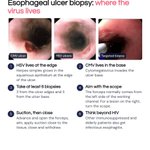
Esophageal ulcer, infection suspected:
HSV lives at the ulcer edge.
CMV lives in the ulcer base.
Take at least 6 biopsies: 3 edge, 3 base. Turn the scope to aim the forceps.
How many do you take?

Full lecture on EndoCollab: endocollab.com/blogs/full-co…

EndoCollab Quiz
Giant gastric folds; initial biopsies were negative.
What is the most likely diagnosis?
a. Ménétrier disease
b. Gastric lymphoma
c. Zollinger-Ellison syndrome
d. Linitis plastica (signet ring cell gastric cancer)
Vote in the reply to see the answer.

Answer in our Telegram quiz. Vote to see it:
t.me/endocollab/65

Looks like a hernia. It is esophagus.
In extensive Barrett's, the distal esophagus is columnar-lined and can show pseudo-folds. The palisade veins of the distal esophagus are now easy to see.
Would you have called this stomach?
Coin lodged in a child's upper esophagus.
1. Grasp tightly, no tissue: rat tooth or alligator forceps
2. Pull slowly, with CO2
3. Stop at the cricopharynx, let the coin dilate the sphincter, then pull
About a minute. Your go-to forceps?
Full lecture on EndoCollab: endocollab.com/blogs/full-co…

USMSTF follow-up after colonoscopy, average-risk adults.
Normal: 10 years. 1-2 tubular adenomas under 10 mm: 7-10 years. 3-4 under 10 mm: 3-5 years. Adenoma 10 mm or larger: 3 years. Piecemeal resection of 20 mm or larger: 6 months.
Assumes a high-quality exam. Not for hereditary syndromes, IBD or CRC history.
Adapted from Gupta et al. 2020, US Multi-Society Task Force.


Small bowel bleeding, 8 mistakes:
1. DBE before repeat EGD/ileocolonoscopy
2. Trusting a negative capsule
3. No CTA if massive, no CT tumor
4. Overt: up to 67% in 48h, 18% day 5
5. Anatomy
6. Anastomosis, LVAD
7. No biopsy
8. APC at 90 or on clips
Which do you see most?

Full lecture on EndoCollab: endocollab.com/blogs/full-co…
