






Un articulo que todo 🫁🔄🫘 debe de leer antes de que acabe el 2026...
1/8 🫁↔️🫘 Lo que ocurre en el ventilador también puede repercutir en el riñón.
Una revisión explora la lesión renal aguda (LRA) asociada a ventilación mecánica y propone integrar mecánica respiratoria, perfusión y congestión. 🧵👇

7/8 🔄 La relación es bidireccional: la LRA puede agravar la disfunción pulmonar por sobrecarga de volumen e inflamación.
🎯 La propuesta: integrar protección pulmonar, perfusión renal y control de congestión, con reevaluación continua.
🧩 Un órgano modifica al otro.
8/8 🤝🫁🫘 Nefrología y medicina crítica necesitan trabajar juntas: integrar ventilación, hemodinamia, balance hídrico y evolución renal.
Compartir decisiones permite abordar al paciente como un sistema conectado.
📚 Gao et al., 2026:
doi.org/10.1186/s40635-026-0…

Mark GN retweeted

New on pulmonarypocus.com: a free, interactive M-mode Lab :pulmonarypocus.com/mmode-lab🫀🫁
Move the M-line and watch the trace change:
EPSS and reduced EF
Mitral stenosis
Lung sliding vs barcode sign
TAPSE and MAPSE
IVC
Any feed back is appreciated reply here or at [email protected]
Mark GN retweeted
New on pulmonarypocus.com: a live Hepatic Vein Doppler Lab 🫀
Watch ECG, CVP and hepatic vein PW Doppler move together as you change RA pressure, TR and rhythm. S > D, S < D, a + S reversal, AF, plus an estimated VExUS grade.
Free, no login: pulmonarypocus.com → Doppler Lab pulmonarypocus.com/cvp-hv
#POCUS #VExUS #MedEd #FOAMed #CriticalCare
Mark GN retweeted

🚨 ¡Convocatoria abierta!
Presenta tus trabajos libres y casos clínicos en el 1er Congreso Internacional de Nefrología Intervencionista.
🏆 ¡Los seleccionados recibirán beca completa para el Congreso + Hands-On Workshop!
📅 Cierre: 15-Dic-26.
🔗 forms.gle/9j5nHbjpS3xEFgn86

Mark GN retweeted

WHERE SHOULD YOU PLACE THE LVOT DOPPLER SAMPLE?
Calculating aortic valve area using the continuity equation requires an accurate LVOT velocity-time integral (VTI).
But a few millimetres can make a difference.
1. Start in the LVOT
Use pulsed-wave Doppler and position the sample volume below the aortic valve.
2. Move toward the valve
Advance the sample volume gradually toward the aortic valve, observing the spectral waveform.
3. Identify the optimal position
As the sample enters the flow acceleration zone, spectral broadening appears. This indicates that the sample is too close to the valve.
4. Move slightly apically
Withdraw the sample until spectral broadening disappears and a clear, laminar LVOT signal is obtained.
Why does this matter?
If the sample is too close to the valve, the measured LVOT VTI may be falsely elevated, causing the calculated aortic valve area to be overestimated.
If it is too far into the ventricle, the measurement may not represent the appropriate LVOT flow velocity.
Clinical pearl: Correct LVOT sample positioning is essential for reliable grading of aortic stenosis.
Reference: ASE recommendations for echocardiographic assessment of aortic stenosis; 2025 ESC/EACTS Guidelines for the Management of Valvular Heart Disease.

Mark GN retweeted

In 1992 Peter Ratcliffe received this rejection from Nature.
Almost 30 years later he won the Nobel Prize for the same discovery.
Don't lose faith in the things you believe in.


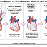
4/5 📉 Los autores proponen:
Carga sostenida → adaptación del VD → dilatación y disfunción → falla derecha y congestión.
⚠️ Es una hipótesis aún no validada longitudinalmente en esta población. No es el destino inevitable de una fístula.
5/5 🩺 ¿Qué nos deja?
Individualizar el acceso y considerar evaluar el VD antes y después de crearlo en pacientes de alto riesgo.
El artículo no demuestra superioridad del catéter. Propone prestar más atención al corazón derecho.
doi.org/10.2215/CJN.00000011…

Mark GN retweeted

Today's Paper of the Day is:
Acute kidney injury in patients with burns
criticalcarereviews.com/late…
Join us to read 1 paper per day and stay up-to-date as we cover the spectrum of critical care across 2026

