

Free online repository & tool for scientific research on phenotypes. Support: @NSF & institutional members. Part of @phoenix_bio. @morphobank@ecoevo.social
- Tweets882
- Following738
- Followers832
- Likes251




ALT Eight moths and one caterpillar from the moth family Psychidae (grey and mottled unless otherwise noted): Lomera boisduvalii (fluffy orange body), Trogonocyttara clandestina, Cebysa leucotelus, Lepidoscia heliochares, Canephora hirsuta (yellow and black striped caterpillar in a case), Lepidoscia confluens (dark brown with broken white stripes), Cryptothelea nigrita (black with triangular wings), an unnamed Iphierga sp. (cream with broken dark brown stripes and an orange head) and Bankesia conspurcatella.


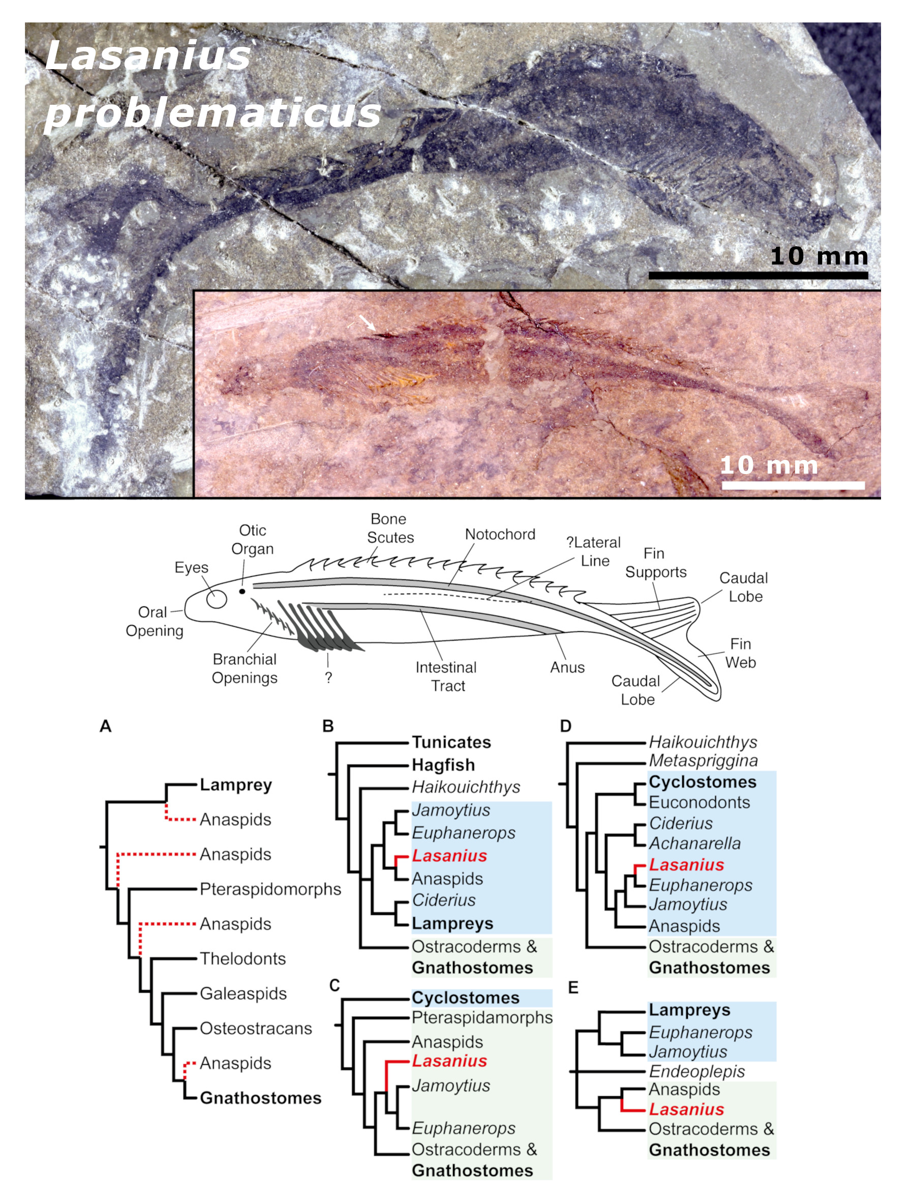
ALT Top: two specimens of Lasanius problematicus in lateral view: Upper photo = NHMUK OR1669 (anterior to right); lower photo = BGS GSE 3989 (anterior to left); both scale bars (in lower right corner of each image) are 10 mm. Middle: stylised sketch in lateral view with main features labelled. Bottom: A selection of the many phylogenetic reconstruction hypotheses. A, proposed positions of the Anaspida (including Lasanius) indicated by a dotted line as: stem-lampreys; the most basal stem-gnathostomes; a group within the ostracoderms; the nearest sister group of gnathostomes. B–E, selected conflicting published hypotheses of Lasanius: B, as stem-lampreys, sister group to the anaspids; C, as stem-lampreys but more closely aligned to Euphanerops than the anaspids; D, as derived stem-gnathostomes outside Anaspida; E, as basal stem-gnathostomes within Anaspida.

ALT Upper part - specimen images on black background: views of the new Hamadasuchus mandible (MNHN-SAM 136) in dorsal (top) and ventral (bottom) view, with features labellled; scale bar 5 cm. Below is a map of the region with a red triangle indicating the locality of La Gara Samani in Algeria (lower left quadrant). Scale bar on the left is 200 km; North upwards.

ALT Top left stratigraphic log of the Kuanynchiao Bed (Hirnantian, Upper Ordovician) indicating the ages, graptolite biozones, levels and lithologies from which brachiopods were collected. Other images are photographs of Eostropheodonta parvicostellata from the Hetaoba section, exhibiting three morphological phenotypes described in the paper, each with a detail sketch highlighting the morphological differences. Bottom left: Ventral mould (NIGP 200426) with a sketch of phenotype A morphology to the right. Top right: Sketch of the phenotype B morphology with an example specimen photograph below: a ventral internal mould showing primitive denticular plates (NIGP 200435). Bottom right: Sketch of phenotype C with an example specimen photograph below: a ventral internal mould (NIGP 200443). All scale bars (in lower left corner of specimen photographs) are 2 mm.

ALT New genus (and combination), Bohemiacinctus havliceki: dorsal view of specimen VK6 (Czech Geological Survey of Prague). Scale bar at bottom right is 2 mm.

ALT Top left: Map of the major Gondwana basins of peninsular India showing the Rewa basin (in grey), other Gondwanan basins in black, and the study area (arrow near top of image). Top middle: Osteoderm from Colossuchus techniensis (IITKGPR392C); scale bar (above image; approximately full length of specimen) is 30 mm. Top right: Anterior view of a middle caudal vertebra (IITKGPR751); scale bar (below, right) is 30 mm. Centre images: the available and well preserved specimens shown in approximate anatomical positions (scale bar on left 1 m); below is a photograph of the thick red mudstone units of the Tiki formation with a person (in blue shirt and white hat) standing approximately one-third from the left of the image. Bottom left: Photograph with interpretative line drawing of a nearly complete mandible (IITKGPR600); scale bar (between two images) is 30 mm. Bottom right: A complete left femur (IITKGPR604) in anterolateral view. Scale bar (below, right) is 60 mm.

ALT Top left: specimen of Conicula striata showing cone like structure with tentacles at the top. Main structure is purple in colour with pale, fine-grained matrix behind. To the right is a line diagram interpreting various features: tentacles in blue/green at the top, a yellow/green pharynx, purple and brown shades indicating the gastric cavity beneath that. Bottom: a reconstruction of Conicula striata as a stem-group Medusozoan with the various features highlighted in similar colours to the diagram above: exterior view on the left, circular cross sections at two points in the middle; interior, cut-away view on the right, with a colour legend indicating the various features on the far right.

ALT Central image: artist's reconstruction of the seabed with conical Palaeoconotuba in the foreground and other representatives of the faunal community (e.g. trilobite in mid-right foreground; Cambrorhytium in the right background, swimming crustacea); sunlight filtering down. Below: summary of the phylogenetic relationships produced in this analysis, as relates to the Cnidaria. On the far left: two images of Palaeoconotuba elongata specimens (ELEL-SJ102087 above and YPLP-13856 (courtesy of Peiyun Cong) below). On the far right: two images of Palaeoconotuba yunnanensis (ELEL-SJ080936A above and ELEL-SJ102086A below).


