

Equation loving Magnetic Resonance Imaging (MRI) scientist and pre-developer at Siemens Healthcare.
- Tweets566
- Following642
- Followers834
- Likes2.3K


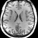




ALT While T1-weighted MPRAGE-based brain morphometry is readily available in clinical routine for adult patients, clinical adoption for children is slow. In particular, normative ranges (i.e., ranges for volumetric values of different brain regions considered healthy) have so far not been available for pediatric patients. Additionally, it would be desirable to have integrated processing that allows simultaneous assessment of morphometry and quantitative values. The authors pursued the idea to implement such a strategy based on a single MP2RAGE acquisition that provides both anatomical information (the MPRAGE-like “uniform” contrast) for morphometry, and quantitative T1 maps.

ALT Data-driven retrospective motion correction can be formulated as a “do-no-harm” strategy. These methods extract the motion information and the motion-free image directly from the k-space data of the imaging scan without requiring additional tracking hardware or disruptive sequence modifications. This ensures that the baseline image quality and contrast will be maintained and that the motion-mitigation strategy only adds value to the exam. Radiologists have the opportunity to directly compare the motion-modeled reconstruction against the original non-corrected images for improved diagnostic utility. Unfortunately, data-driven motion correction is computationally demanding because it often requires the inversion of a large and sophisticated nonlinear physics model that describes the effect of motion on the acquired k-space data. The scout accelerated motion estimation and reduction (SAMER) algorithm will reduce the computational burden of motion estimation.


ALT The need for increased medical autonomy is not unique to space travellers. Remote and medically isolated populations experience many of the same barriers found in space. These may include: • limited medical equipment and resources • communication delays or difficulties • lack of access to medical expertise • limited possibility of emergency evacuations These challenges make it more difficult for people living and working in remote communities to get the care they need. Doctors in these communities could use improved telemedicine technologies and other tools to provide a range of care, from routine exams to serious emergency treatment. Autonomous medical care could help the health professionals in these distant areas diagnose and treat patients right in their communities.


ALT The field of diffusion MRI is much richer than diffusion tensor imaging (DTI). There is significant value in retrieving specific information about features such as cellular density, morphology, characteristic sizes, myelination, etc. This is the challenge that the field of microstructural MRI has taken on, with remarkable progress and success stories over the past few years. Diffusion kurtosis imaging (DKI) can provide complementary information to DTI about tissue microstructure. For example, in voxel-based or ROI-based analyses, kurtosis metrics can identify differences between patients and controls in brain regions where DTI metrics do not. DKI is thus used to improve the detection of brain changes during, for instance, development, aging, early psychosis and schizophrenia, cognitive impairment and Alzheimer’s disease, multiple sclerosis, glioma, stroke, traumatic brain injury, and prostate and other body applications.
ALT The field of diffusion MRI is much richer than diffusion tensor imaging (DTI). There is significant value in retrieving specific information about features such as cellular density, morphology, characteristic sizes, myelination, etc. This is the challenge that the field of microstructural MRI has taken on, with remarkable progress and success stories over the past few years. Diffusion kurtosis imaging (DKI) can provide complementary information to DTI about tissue microstructure. For example, in voxel-based or ROI-based analyses, kurtosis metrics can identify differences between patients and controls in brain regions where DTI metrics do not. DKI is thus used to improve the detection of brain changes during, for instance, development, aging, early psychosis and schizophrenia, cognitive impairment and Alzheimer’s disease, multiple sclerosis, glioma, stroke, traumatic brain injury, and prostate and other body applications.


ALT The potential impact of deep learning-enhanced MRI sequences in the future is considerable. Shorter scan times can enhance the patient experience and allow for higher daily patient volumes, thus broadening access to MRI services. Furthermore, these advanced MRI sequences can also elevate image quality, potentially enabling more precise diagnosis and more effective treatment planning. This improvement in diagnostic accuracy and efficiency could lead to enhanced patient care and possibly lower healthcare expenditure. Grasping the utility and advantages of deep learning in MRI, particularly in speeding up scans and elevating image quality, is therefore vital. The authors provide a comprehensive overview of the daily applications that are already part of their clinical routine.

ALT Undoubtedly, MRI is the cornerstone for imaging traumatic and degenerative knee conditions. Nevertheless, challenges persist in diagnosing partial cruciate ligament injuries, differentiating meniscus tears from signal alterations in normal anatomy, and identifying early cartilage clefts. The advent of ultra-high-field (UHF) MRI at 7T holds promise for addressing these challenges. Using tailored imaging sequences in an ultra-high-field setting notably enhances resolution, providing greater confidence for the anatomical assessment of the knee. The incorporation of 3D sequences further addresses common pitfalls encountered in routine scans at 3T and 1.5T. Optimizing the protocol with the latest AI developments, such as the Deep Resolve options, facilitates the acquisition of high-quality sequences within a reasonable time frame.


ALT The BioMatrix Beat Sensor is an innovative technology that can acquire biological information including cardiac activity and diaphragmatic motion without placing ECG leads or respiratory belts. Therefore, this technique can simplify patient preparation, reduce examination time in cardiac MRI examinations, and improve the patient experience during the scan. In this article, the authors demonstrate the advantages of Beat Sensor triggering in arrhythmic patients and in difficult hearts.

ALT Embracing the spirit of this year's RSNA theme, "Leading through change," Professor Ulrike Attenberger (University Hospital Bonn, Germany) explores the pivotal role of digitalization and AI in creating efficient, patient-centric healthcare systems. The articles in this issue delve into AI-based image reconstruction algorithms (DeepResolve); optimized brachial plexus MRI; workflow support with 3D Whole Heart; MR Elastography; MR-guided thermoablation; or ways to achieve standardized high image quality in Neurological MRI. To name just a few of the inspiring topics. We extend our heartfelt gratitude to the authors and to all those who contributed to the successful realization of this edition of MAGNETOM Flash. Their dedication, expertise, and collaborative spirit have been instrumental in shaping this publication.

ALT MRN comprises a specialized set of techniques focused on imaging peripheral nerves, which can be challenging given their small size and non-linear course throughout the body. When utilized in parallel with electrodiagnostic testing, #MRN not only facilitates accurate localization of nerve injuries but also offers a comprehensive assessment of both the injured nerve and the endorgan muscle. MRN of the brachial plexus presents unique challenges due to inherent field inhomogeneities related to the neck/shoulder curvature and proximity to the lungs. The article covers coil configuration, protocol optimization, respiratory triggering and an outlook on MR Neurography in the future.


