




Vladyslav Kavalerchyk 🇺🇦 🇩🇪 retweeted

#echofirst case report about functional TR non atrial nor ventricular
👉 academic.oup.com/ehjcr/advan…
@dr_benoy_n_shah @echo_batman @echo_stepbystep @StellEkaterina

Vladyslav Kavalerchyk 🇺🇦 🇩🇪 retweeted

A multicentre study published in European Heart Journal – Imaging Methods and Practice evaluated the relationship between ventricular fibrosis and recovery of LV function after catheter ablation for atrial fibrillation.
The study included 134 patients with AF and LV systolic dysfunction. Following ablation, 101 patients (75.4%) achieved meaningful LV recovery.
🔹 Ventricular late gadolinium enhancement (LGE) was more common among patients without recovery: 69.7% vs 46.5%.
🔹 Patients who did not recover had nearly twice the LGE burden compared with responders: 6.6% vs 3.3% (p<0.001).
🔹 Each 1% increase in LGE burden was associated with a lower likelihood of LV recovery (RR 0.96; 95% CI 0.92–0.99; p=0.036).
🔹 The visual pattern and distribution of LGE were not associated with recovery.
🔗 doi.org/10.1093/ehjimp/qyag1…
@EHJIMPEiC @VikashJaiswalMD
#AtrialFibrillation #CatheterAblation #CardiacMRI #LateGadoliniumEnhancement #HeartFailure #LeftVentricularFunction #MyocardialFibrosis #Cardiology #CardiacImaging #RhythmControl #Electrophysiology #CardioEd #CardioX

Vladyslav Kavalerchyk 🇺🇦 🇩🇪 retweeted
Papillary fibroelastoma is a rare primary cardiac tumour with benign histology. Despite its benign nature, it can lead to serious complications, including thromboembolism, stroke, myocardial infarction, and dynamic coronary obstruction.
This review at #EHJ-IMP highlights how multimodality imaging supports diagnosis and clinical management:
🔹 Transthoracic echocardiography (TTE) serves as an important initial imaging test.
🔹 Transoesophageal echocardiography (TEE) provides improved spatial and temporal resolution, helping define tumour mobility, attachment, stalk morphology, and relationships with nearby structures.
🔹 Cardiac CT offers detailed anatomical information and helps assess the tumour’s relationship with the cardiac valves, coronary arteries, and surrounding anatomy.
🔹 Cardiac MRI contributes to tissue characterisation and may help differentiate papillary fibroelastoma from other cardiac masses.
🔗 doi.org/10.1093/ehjimp/qyag1…
@VikashJaiswalMD @EHJIMPEiC @JGrapsa
#EHJImaging #Cardiology #CardiacImaging #MultimodalityImaging #Echocardiography #CardiacCT #CardiacMRI #PapillaryFibroelastoma #CardioEd #CardioTwitter.

Vladyslav Kavalerchyk 🇺🇦 🇩🇪 retweeted

Rare congenital anomaly(I’ve never seen)
rudimentary aortic chordae tendineae #echofirst
seen as ruptured fibrous strand attached to RCC free edge w partial flail causing severe aortic regurgitation OR confirmed bit.ly/3ULiWIb
Vladyslav Kavalerchyk 🇺🇦 🇩🇪 retweeted

A mitral-aortic intervalvular fibrosa pseudoaneurysm is a rare complication after cardiac surgery.
This report describes the diagnosis & surgical management of an MAIVF pseudoaneurysm in a 3-year-old after the resection of a recurrent subaortic membrane. bit.ly/4elxsgo
Vladyslav Kavalerchyk 🇺🇦 🇩🇪 retweeted

A routine day in the Echolab 😎 for 4D echo lovers @NMerke @echo_stepbystep @denisamuraru @fiore_corrado @drozgeozden @DrHaniMahmoud @DonalErwan @lpbadano @GEHealthCare
Vladyslav Kavalerchyk 🇺🇦 🇩🇪 retweeted

A defining study on evaluating MR with Echocardiography & impact on @ASE360 Guidelines. @ACCinTouch @SCMRorg

🎯 Less is more in mitral regurgitation assessment
In 177 patients undergoing near-simultaneous Echo & CMR, just 2 ASE criteria identified significant MR with 91–95% specificity.
🫀 Smarter integration, less quantification, greater diagnostic confidence
doi.org/10.1016/j.jcmg.2026.…

Vladyslav Kavalerchyk 🇺🇦 🇩🇪 retweeted

#JADEL
New in @CASEfromASE... large RA mass.
Given the imaging findings shown, what is the most likely pathology?
A. Angiosarcoma
B. Myxoma
C. Lipoma
D. Rhabdomyoma
E. Thrombus
Follow thread for pathology... (but, answer before looking)? 🤔
@ASE360


Vladyslav Kavalerchyk 🇺🇦 🇩🇪 retweeted

New #JACC State-of-the-Art Review: Clinical Considerations in Management of Hypertrophic Cardiomyopathy in Pregnancy
Read here: bit.ly/4hXr6pN
#CardioX #HCM

Vladyslav Kavalerchyk 🇺🇦 🇩🇪 retweeted

🫀 How are you applying the 2025 ESC/EACTS guidelines in aortic stenosis?
Share your experience with multimodality imaging, clinical challenges and educational needs.
Your voice matters!
🔗 lnkd.in/e6fiYNZU
#AorticStenosis #EACVI

Vladyslav Kavalerchyk 🇺🇦 🇩🇪 retweeted

🎯 Less is more in mitral regurgitation assessment
In 177 patients undergoing near-simultaneous Echo & CMR, just 2 ASE criteria identified significant MR with 91–95% specificity.
🫀 Smarter integration, less quantification, greater diagnostic confidence
doi.org/10.1016/j.jcmg.2026.…
Vladyslav Kavalerchyk 🇺🇦 🇩🇪 retweeted

Not Every Calcified Aortic Valve Is Rheumatic!
Today, one of my trainees described mild focal calcification of the aortic valve as “rheumatic involvement.”
It sparked a great teaching discussion!
How often does rheumatic heart disease involve the aortic valve in isolation?
✅Isolated aortic involvement accounts for <8% of rheumatic heart disease.
✅ Isolated rheumatic aortic stenosis is even rarer (~2%).
✅ Rheumatic aortic regurgitation is more common than isolated rheumatic AS.
✅ Most rheumatic aortic valve disease occurs alongside mitral valve involvement.
🔍 What should we look for on echo?
🫀Commissural fusion — a hallmark of chronic rheumatic valvular disease.
🫀 Leaflet thickening, particularly at the free edges.
🫀 Restricted leaflet mobility.
🫀 Progressive fibrosis and calcification.
Calcification alone does not establish rheumatic etiology.
Before calling an aortic valve rheumatic, look at the commissures—and don’t forget the mitral valve!
Sometimes, the most important imaging finding is what isn’t there.
#EchoFirst #CVImaging #RheumaticHeartDisease #CardioTea
PS found this really nice schematic on insta- if anyone knows the author on X please tag him. I am not able to find him here!
Vladyslav Kavalerchyk 🇺🇦 🇩🇪 retweeted
A new report in #EHJIMP highlights a rare but potentially life-threatening complication of mitral transcatheter edge-to-edge repair (M-TEER): right-to-left shunting through an iatrogenic atrial septal defect (iASD).
🔍 Key findings from 444 patients undergoing successful M-TEER:
▪️ 6 patients (1.3%) required emergency iASD closure for acute deoxygenation.
▪️ All had severe tricuspid regurgitation, right ventricular (RV) dilatation, and impaired RV function.
▪️ In 5 of 6 patients, right atrial pressure exceeded left atrial pressure after M-TEER.
▪️ RV function was substantially impaired, with lower TAPSE (15 vs. 19 mm), fractional area change (27% vs. 43%), and RV free-wall strain (−13% vs. −22%).
▪️ Echocardiography underestimated invasively measured systolic pulmonary artery pressure (25.8 vs. 40.3 mmHg; P = 0.035).
▪️ Oxygen saturation decreased by 3–8%; one patient developed cardiogenic shock.
Read the article: doi.org/10.1093/ehjimp/qyag1…
@VikashJaiswalMD @EHJIMPEiC
#Cardiology #StructuralHeart #MTEER #MitralRegurgitation #Echocardiography #RightVentricle #CardiacImaging #Cardiotwitter

Vladyslav Kavalerchyk 🇺🇦 🇩🇪 retweeted
Can clinically manifest cardiac sarcoidosis occur in the absence of LGE on CMR?
A new study published in #EHJ-IMP provides evidence that clinically significant, metabolically active cardiac sarcoidosis may occur despite the absence of late gadolinium enhancement (LGE) on cardiac magnetic resonance (CMR).
🔬 Key findings
• Among 46 treatment-naïve patients with clinically manifest, FDG-avid cardiac sarcoidosis, 4 (8.7%) had no detectable LGE on CMR.
• All 4 LGE-negative patients presented with advanced conduction system disease; 3 had complete heart block.
• Left ventricular ejection fraction was higher in LGE-negative than in LGE-positive patients (70% vs. 50%; P = 0.010).
• Myocardial inflammatory burden, assessed by LV SUVmax on FDG-PET, was lower in LGE-negative patients (3.9 ± 0.9 vs. 9.6 ± 5.2; P = 0.037).
• Three of the 4 LGE-negative patients received immunosuppressive therapy.
📄 Read the full article: doi.org/10.1093/ehjimp/qyag1…
@EHJIMPEiC @VikashJaiswalMD
🗣️#CardioTwitter #Cardiology #CardiacSarcoidosis #Sarcoidosis #WhyCMR #CVImaging #NuclearCardiology #FDGPET #EPeeps #HeartBlock #Arrhythmia #HeartFailure #MedEd #EHJIMP

Vladyslav Kavalerchyk 🇺🇦 🇩🇪 retweeted
#echofirst 3D Compilation
Teasing #3DEchoBerlin
22.01.- 24.01.2027 Advanced Echocardiography Workshop
Sunday 3D Hands on with GE & Philips (TomTec)
👉lnkd.in/eWskntkH
👉 lnkd.in/eWskntkH


Vladyslav Kavalerchyk 🇺🇦 🇩🇪 retweeted
🫀 3D Advanced Echo Workshop | 22–24 Jan 2027, Berlin
#echofirst
International experts, hybrid format. 3D focus, but not only.
Friday's Basics session alone: physics by images, how to do 3D, contrast in unexpected ways, live knobology on GE & Philips real 3D cases. And that's just the start.
@echo_batman @alexsfelixecho @papadocardio @fiore_corrado
And many more to come
👉 shorturl.at/VzVzf
👉 shorturl.at/rdvQp


🚀 ICE is transforming cardiac interventions!
@DonalErwan & @AugustinCoisne highlight the shift from procedural guidance to precision intervention.
🔹 Real-time 3D/4D imaging
🔹 Safer, smarter workflows
🔹 AI-assisted navigation
☑️doi.org/10.1093/ehjci/jeag21…
@EHJCVIEiC

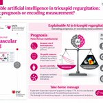
🚨 Can AI decode TR prognosis—or merely encode measurement errors?
@lpbadano & Penso challenge PISA-derived EROA, Doppler-estimated pressures & missing quantitative RV assessment.
AI 🤖 cannot correct flawed measurements!
☑️doi.org/10.1093/ehjci/jeag19…
@EHJCVIEiC @EACVIPresident
