

PGY-4 pathology resident, interested in gyn path and soft tissue path.
United States
Joined October 2018
- Tweets948
- Following847
- Followers775
- Likes1.4K




Wangpan “Jackson” Shi,MD retweeted

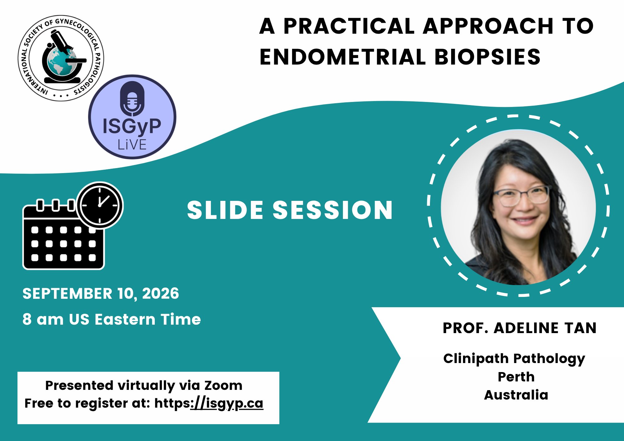
Announcing a free @ISGynP sponsored live virtual slide session on Sep 10 @ 8 AM US ET by Dr. Tan - "A Practical Approach to Endometrial Biopsies". Register now @ isgyp.ca
#gynpath #gynaepath #PathTwitter
Wangpan “Jackson” Shi,MD retweeted

Excited to share our @Pathoutlines article on OGT-rearranged mesenchymal neoplasm has been published online. Grateful to everyone for their help/contribution!
Check it out
👇🏻👇🏻👇🏻👇🏻
pathologyoutlines.com/topic/…
3 things to remember about OGT-rearranged mesenchymal neoplasm (also known as myoepthelioma-like hyalinizing epithelioid tumor, MHET):
💎 Well circumscribed, multinodular, composed of bland epithelioid to spindled cells set in a 2 toned stroma with myxochondroid (blue) and acellular hyalinized (pink) areas
💎 Signet ring-like cells with cytoplasmic processes engulfing extracellular hyaline globular material
💎 No IHC evidence of myoepithelial diff (➖ve for AE1/ AE3, SOX10, S100P, GFAP)
Consistently ➕ve for CD34
🌟Bonus info: Recurrent fusions of OGT and members of FOXO, most commonly FOXO3, FOXO1, FOXO4
#PathTwitter #BSTPath

ALT Two-toned stroma: hyaline and myxoid areas

ALT Signet ring-like cells
Wangpan “Jackson” Shi,MD retweeted
Don't miss this great opportunity to learn #BSTpath from fantastic lecturers, with 150 cases to preview & a structured approach to this beautiful subspecialty.
Been attending since 2024 and it never disappointed me.
On-site & virtual! Maybe at some point I'll be able to attend in-person 🥺
#pathresidents #pathtwitter

Registration is now officially open for our third annual Cleveland Clinic Soft Tissue Pathology Course!! May 14-17, 2026. Live and VIRTUAL options!! 150 brand new cases!! #pathology #softtissue #sarcoma #bstpath #PathTwitter
Link:
na01.safelinks.protection.ou…
@Williamson_SR @smlungpathguy @JMGardnerMD @MirunaPopescu13 @cebulka26 @evemariecrane @ClevelandClinic @CleClinicNews @CCFPathRes

Wangpan “Jackson” Shi,MD retweeted

Three variations on seminoma #testiscancer #gupath
#1 - seminoma w/transitions to embryonal carcinoma - differences in cytoplasm, nuclear contour, & nucleoli - both OCT-4(+); diffuse membranous labeling for CD117/D2-40 (seminoma) vs. CD30 (EC) = helpful differential stains


Wangpan “Jackson” Shi,MD retweeted
28 y/o M w/ 12 cm retroperitoneal mass attached to and infiltrating bowel wall.
What are your thoughts? 🔬
Credits🙏🏼: thepathologist.com/exercise-… (check out free WSI here & many others! Great learning resource)!


No definite answer but more of sharing the findings. EMB with hormone therapy after initial dx of atypical hyperplasia/EIN. How would you sign out with these findings?


Elderly female with an 6.2 cm ovarian mass. Received chemo before the surgery due to large effusion.
What’s your morphology impression?
#PathTwitter

Sorry, this isn’t a perfect case to prove the point because no immunostain is available, as it’s from a very old case study set. It was diagnosed as clear cell carcinoma of the ovary, and the unusually ugly appearance is probably due to chemotherapy. However, we definitely need to perform additional stains in real life.
Mass from heart. What's your impression by morphology? #PathTwitter


Wangpan “Jackson” Shi,MD retweeted

Thyroid-Benign tumors
-Thyroid follicular nodular disease is the new name for what clinically is known as multinodular goiter.
"Multifocal non-inflammatory benign proliferation of thyroid follicular cells that results in multiple clonal and non-clonal nodules with highly variable architecture"
Usual terms used to sign out this is multinodular hyperplasia/adenomatous hyperplasia/nodular goiter
Question for the experts? Do you use this terminology to sign out cases?
Pic from Dr. Nose presentation at CCP2024 #pathology #pathologists #PathX #PathTwitter #pathresidents #pathfellows

Wangpan “Jackson” Shi,MD retweeted

🔬Dx: Microcystic/reticular schwannoma
Has a predilection for visceral locations
See references by Dr Fletcher and Dr @RunjanChetty
ncbi.nlm.nih.gov/m/pubmed/18…
ncbi.nlm.nih.gov/m/pubmed/20…


Wangpan “Jackson” Shi,MD retweeted

S100+++
MICROCYSTIC/RETICULAR SCHWANNOMA
Case courtesy: Liegl, B., Bodo, K., Martin, D., Tsybrovskyy, O., Lackner, K. and Beham, A. (2011), Microcystic/reticular schwannoma of the pancreas: A potential diagnostic pitfall. Pathology International, 61: 88-92.


Thigh mass in an elder patient. Are you worried about sarcoma? #PathTwitter

Right parapharyngeal mass. What category of tumor? #PathTwitter


AR+ (as shown in the figures) was observed in the ductal cells. Although no infiltrative or invasive pattern was noted on low power, the positive AR and cytologic atypia in the luminal cells led to the decision to classify it as apocrine intraductal carcinoma ex PA. HER2 is also positive in luminal cells, and Ki-67 is approximately 5%.


A frozen section from parathyroid. How would you report it? #PathTwitter


