
MD; former Chief Resident IM Baptist Hosp Beaumont,TX, Guitarist, Singer, Swimmer,
New Orleans, Louisiana
Joined March 2015
- Tweets994
- Following3.1K
- Followers834
- Likes1.3K
📷 Is it going to be considered new risk factor for coronary atherosclerosis ? Ear Lobe Crease - Frank's sign
Read our new publication
pubmed.ncbi.nlm.nih.gov/4293…

Glad to share the latest publication in the Journal of American Heart Association. Thank you, my mentors and co-authors.
ahajournals.org/doi/10.1161/…
@JAHA_AHA @AHAScience @ACCinTouch @Cardiology

Excited to share the book chapter on obesity and the Obesity Paradox in Heart Failure in Encyclopedia of Cardiovascular Research and Medicine(2e) (Second Edition)
sciencedirect.com/science/ch…

Ankit Vyas retweeted
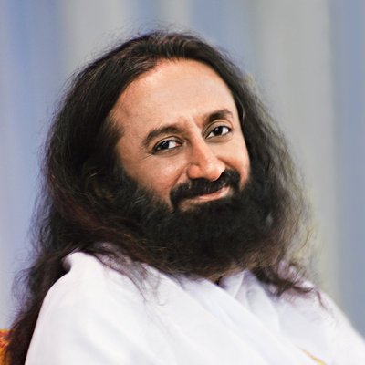
The busier you are in life, the more important it is to meditate. Meditation is getting back to that serenity which is your original nature.
Ankit Vyas retweeted

M-Mode Echocardiography Explained
This figure demonstrates M-mode measurements from the parasternal long-axis (PLAX) view, crucial for assessing valvular & ventricular function:
🔹 Aortic Valve Motion (Top Panel)
- The right coronary cusp (RCC) and non-coronary cusp (NCC) move apart in systole (valve opens) and come together in diastole (valve closes).
🔹 Mitral Valve Motion (Middle Panel)
- The anterior mitral leaflet (AML) and posterior mitral leaflet (PML) exhibit characteristic movements:
- Diastole: AML moves towards the septum (E wave) due to rapid LV filling, then slightly back (A wave) due to atrial contraction.
- Systole: Leaflets close tightly, preventing regurgitation.
🔹 Left Ventricular Dimensions (Bottom Panel)
- End-diastolic diameter (EDD): Largest LV dimension measured before systole.
- End-systolic diameter (ESD): Smallest LV dimension after contraction.
- These values help calculate LV function (e.g., fractional shortening & ejection fraction).
📌 Courtesy of Bernard E. Bulwer, MD, FASE
#Echocardiography #Cardiology #Mmode

ALT Illustration of M-mode echocardiography from the parasternal long-axis (PLAX) view. The aortic valve panel shows RCC and NCC separation during systole and closure during diastole. The mitral valve panel displays anterior and posterior mitral leaflet movements (E and A waves). The left ventricle panel highlights LV cavity dimensions, with EDD and ESD measured to assess function.


medicalxpress.com/news/2025-… Before you give drinks or juice to your kids or yourself, think twice. Tufts University, USA, published research. It's worth reading. Healthy Heart!

How to get into Cardiology Fellowship - American College of Cardiology webinar. My insights from 00:06:00 - 00:14:00
youtu.be/r04NYGz2Cso
Ankit Vyas retweeted

In 25 yrs in Cath lab .. for the first time did procedure without lead apron .Rampart shield (radiation protection ) will make our spine and life better
@drpkhazra @DrSaritaRao2 @Hragy @NishithChandra @mirvatalasnag @mmamas1973 @tavrkapadia @mandeep_mayo @mathur_a @SachinGoelMD


Ankit Vyas retweeted

Day 2 #24FellowsAPP conference kicking off hot with an expert, multispecialty panel of speakers on #PAD 🔥🦵 teaching to save lives and limbs! @AaronAdayMD @vivianbishay @DrDrewKleinPHI @wellsbryanj @adityasharmamd @YSCastroMD @OAlabiMD @SVM_tweets – at Atlanta, GA


Ankit Vyas retweeted

Medical management for #riskfactor modification is crucial in #PAD!#Revascularization may have a role in symptomatic pts & to reduce tissue loss in CLTI
#SaveLimbs #SaveLives @PVDCouncil
@adityasharmamd @PiedmontHealth @DrDrewKleinPHI
@wellsbryanj @AaronAdayMD @heatherlgornik


Excited to share our latest publication on cardiac arrest outcomes in people with obesity and Bariatric surgery. Please read and give comments. Thank you
sciencedirect.com/science/ar…
@OchsnerCVFellow @OchsnerHealth @Skhatib55 @Stent4U @SCAI @AHAScience @ACCinTouch @LAACC_WIC @IJCCRP
Ankit Vyas retweeted


NEW EP on EP episode: @EPrystowskyonEP is joined by @DanielPMorin to discuss Mobile Technology in AF Management. See the full interview here #EPeeps ➡️ bit.ly/3v7K0En